Klatskin's tumor
1. perihilar bile duct tumor,位於肝管分枝(bifurcation of hepatic duct)
2. Usually presents between ages 50-70 but can present earlier in pts with primary sclerosing cholangitis (PSC) and in pts with choledochal cysts. Slightly higher incidence in men.
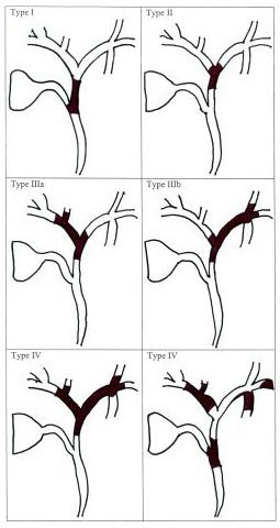
3. Bismuth classification:
a)
type
1:腫瘤在左右肝管的匯合區以下(below confluence of
hepatic duct)
b) type
2:腫瘤到達匯合區(reaching confluence)
c) type
3:阻塞總肝管(common hepatic duct)和右肝管(IIIa)或左肝管(IIIb)
d) type
4:腫瘤是多中心的(multicentric)或侵犯匯合區以及左右肝管

4. Risk factor
a) Hepatolithiasia : 5-10%發生膽管癌
b) Parasitic infections: Liver flukes (Clonorchis and Opisthorchis) are associated with intrahepatic cholangiocarcinoma
c) Choledochal cysts
d) Sclerosing cholangitis
e) Oral contraceptives
5. Pathology: 多為Adenocarcinoma
外觀上可分為:
a) local or nodular: 大約2公分, annular, constricting, 灰白色
b)
diffuse: 整個膽管廣泛地增厚; 從肝門到肝臟
c)
papillary: 突出到膽管內腔; multple, diffuse
6. Clinical symptoms
obstructive jaundice: 90%
RUQ pain: 30~50%
severe、persistent pruritus: 60%
body weight loss: 30~50%
fever: 20%
hepatomegaly: 25~40%
Classic triad for hepatobiliary or pancreatic cancer: cholestasis, abdl pain, weight loss.
7. Diagnosis
a. Lab data:
Bilirubin↑(> 10 mg/dl), Alk-p↑(2~10 X), GGT↑, INR↑, biliary CEA ↑
If CEA> >5.2 ng/mL + CA 19-9 >180 U/mL=> sensitivity 100%
b. Ultrasound: segmental dilatation or nonunion of R and L ducts, polypoid intraluminal masses, nodular smooth masses with mural thickening. Should do Doppler, as this is helpful to assess vascular invasion (unresectable)
c. CT: A contracted gallbladder is more typical of a Klatskin tumor whereas a dilated GB is suggestive of a common bile duct tumor.
d. Cholangiography (ERCP or PTC): ERCP has benefit of obtaining cells for biopsy
e. MRCP: similar to CT, cholangiography, and angiography combined. Early studies show PPV 86% and NPV 98%
8. Staging
|
Stage 0 |
Tis |
原位癌 |
|
|
Stage I |
IA |
T1 |
組織學上腫瘤侷限在膽管(bile duct) |
|
IB |
T2 |
腫瘤侵犯超出膽管壁(beyond the wall of bile duct) |
|
|
Stage II |
IIA |
T3 |
腫瘤侵犯肝臟、膽囊、胰臟, 和/或 門靜脈(左或右)或肝動脈(左或右)的單側分枝 |
|
IIB |
N1 |
局部淋巴轉移 |
|
|
Stage III |
T4 |
腫瘤侵犯下列任一: 兩側的主要門靜脈或它的分枝、總肝動脈(common hepatic artery)、或其它鄰近構造, 例如大腸、胃、十二指腸、或腹壁 |
|
|
Stage IV |
M1 |
meta |
|
9. Prognosis
a. resectable: 5年存活率20%
b. unresectable: 中位存活期5個月
10. Treatment
a. 手術:
resectable:先做choledochoscopy查看腫瘤範圍。
(i) Upper: 包括bilateral hepatic duct, confluence, common hepatic duct
=> resectable小於20%, 所以常做T-tube引流
(ii) Middle: 包括從cystic duct到pancreas這一段的總膽管(CBD) => When possible, excision & duct-enteric
biliary bypass
=> If necessary, partial hepatic resection & cholangiojejunostomy
(iii) Intrapancreatic portion of CBD =>
Pancreaticoduodenectomy
unresectable or metastatic:
(i) 手術減壓(surgical decompression)
(ii) 非手術減壓(non-surgical
decompression)的方法包括: T-tube, endoscopic
papillotomy, biliary stent, bolloon dilation, endoprosthesis insertion


 留言列表
留言列表


